Retroperitoneal Liposarcoma - A Breakdown Of Its Effects And Treatments
An extremely rare tumor called retroperitoneal liposarcoma (RLS) is known for its vast spectrum of physiological features. As a result of its size and depth, it is difficult to cure. Patients with high-grade RLS who undergo surgery are more likely to suffer from locally recurrent disease, which is the leading cause of death for most patients.
Author:James PierceReviewer:Karan EmerySep 17, 20221 Shares160 Views
An extremely rare tumor called retroperitoneal liposarcoma(RLS) is known for its vast spectrum of physiological features.
As a result of its size and depth, it is difficult to cure.
Patients with high-grade RLS who undergo surgery are more likely to suffer from locally recurrent disease, which is the leading cause of death for most patients.
Recurrence is common, even after a removal that has been deemed "grossly complete," necessitating patients to be monitored for long periods of time, often indefinitely.
Although liposarcoma is a rare kind of cancer, it can be fatal if left untreated. Liposarcoma is a form of soft tissue sarcoma that occurs when cancer cells invade fat cells.
Anywhere in the body, fat cells can produce liposarcoma, but the great majority of cases occur in the leg or abdominal muscles.
While liposarcoma can strike adults of any age, it most usually strikes the elderly.
However, unless the tumor has grown large enough to squeeze nearby organs, retroperitoneal liposarcoma usually causes no symptoms.
The illness is frequently misdiagnosed due to its rarity and the absence of any symptoms.
The Anatomy Of The Retroperitoneum
The retroperitoneum (RP) and preperitoneum make up the extraperitoneal area. A portion of the pancreas, part of the liver, and the transverse colon are all located in the anterior region of this area. RP space is defined by these peritoneal extensions. Peritoneal extensions also anchor a section of the duodenum.
The perirenal space, the (anterior and posterior) pararenal spaces, and other important structures make up the retroperitoneum (RP).
A number of organs and systems can be found in the retroperitoneal space (which includes the pancreas, kidneys, and adrenals), the larger abdominal veins, lymphatics, six major nerves, and the autonomic (sympathetic) lumbar chains, as well as the connective tissue.
PRTs, or primary retroperitoneal tumors, are tumors that begin in the RP space rather than in the corresponding organs of the retroperitoneum.

Soft Tissue Sarcomas | FAQ with Dr. Adam Levin
Natural History Of Liposarcoma
Soft tissue sarcoma (STS) incidence in Europe is from 4.5 to 5.0 per 100,000 people each year. Adult malignant tumors have an incidence of this type that is more than one percent. Adult STS make up 10–15% of the total RP population. A liposarcoma is thought to be the cause of 20% of STS sarcomas and 50% of RP sarcomas.
A wide range of possible results can be expected from STS. There are a variety of characteristics that influence how STS behaves and how it will affect a person, including their age, the tumor's location and depth, size, resectability, histology and grade, the existence of nodal disease, and whether or not distant metastases are present (DM).
RPS, a kind of soft tissue sarcoma, rarely spreads to other parts of the body, with only 10% of patients showing signs of metastatic disease when they first arrive at the clinic. When these tumors spread to other organs, they are usually seen in the lungs or liver. DM has the potential to impact the prognosis of any STS.
RPS patients with diabetes have a 20–25 percent mortality rate and a median survival duration of 13 months. Depending on the severity of the tumor, STS and RPS DM are used. Because most RPS are of low quality, DMs are quite rare.
Concerns about recurrence and local control are at the top of the list. Large, anatomically complex RPS are notoriously difficult to remove surgically because of their proximity to so many important structures.
Some Predisposing Factors
There is no known cause for STS or RPS. There are genetic defects and exposure to radiation or chemicals that can increase the risk of cancer. Radiation has a greater effect on genetically susceptible patients.
There is an estimated 5% chance of developing STS in the general population and notably in children (17–19) when CT-scanning and high-dose ionizing radiation are used to treat diseases such as breast cancer, lymphoma, and pediatric cancers.
External beam radiation therapy's effect on the development of STS after treatment is not understood. External beam radiation therapy (EBRT)-associated fibrosarcomas, osteogenic sarcomas, angiosarcomas, leiomyosarcomas, and undifferentiated pleomorphic sarcomas are among the most common radiation-associated STS that are high grade or poorly differentiated and located at the radiation field's edge.
Chemicals including phenoxyacetic acid, thorium bromide (Thorrast), vinyl chloride (Vinyl), arsenic (Asbestos), and androgenic-anabolic steroids (AAS) have been linked to STS in the past, but this is mainly a historical issue.

Dr. George Demetri on Liposarcoma | Dana-Farber Cancer Institute
The Histology And Molecular Biology Of Retroperitoneal Liposarcoma
Histological classifications of liposarcomas have undergone radical changes in the last few decades as a result of discoveries in molecular genetics. Liposarcoma is classified by the World Health Organization into four distinct subtypes according to how well or poorly differentiated, dedifferentiated, or pleomorphic it is.
Sclerosing/inflammatory/spindle cell types of these include lipoma-like (or adipocytic) A wide range of physical and genetic differences can be identified between the various sub-groups. "Mixed or combination liposarcoma" has been deleted from the 2013 WHO classification because consensus has been established that these rare occurrences appear to be (variants of) dedifferentiated liposarcoma.
There are many similarities between atypical lipomatous tumors and well-differentiated liposarcoma; therefore, they should be handled as synonyms.
For patients with an atypical lipomatous tumor, it is possible to employ the tumor as a therapy option. Tumor-free surgical margins greater than 2 cm in the RP are extremely rare.
Recurrence is common even if there is no dedifferentiation or metastasis. In a few rare cases, lipoid tumors are substituted for lipomatous tumors. Histopathology can be used to identify lipomatous tumors. As a result, liposarcoma's histological subtype is a key prognostic predictor, highlighting the importance of subclassification.
Types Of Treatment For Retroperitoneal Liposarcoma
Surgery
The surgical excision of retroperitoneal liposarcoma remains the most crucial component of treatment, and the highest chance for cure occurs at the time of the first surgery. Preoperative imaging and intraoperative results should lead to a complete excision of the tumor, minimizing marginality, and an en-bloc resection of all possibly implicated tissues is desirable.
An abdominal wall, paravertebral muscles, or psoas excision in one piece is frequently required for macroscopically complete surgery. Given the location and size of these tumors and the lack of a consistent technique for processing the specimen, despite continued efforts, no compelling approach has been discovered to assess the adequacy of surgical margins.
Because it lowers marginality and positive margins, an extended surgical approach has a lower recurrence rate than simple excisions.
Operational planning often incorporates a functional evaluation of key organs, such as the kidneys' individual performance. Bilateral renal involvement, encasement of the superior mesenteric artery, celiac axis, and porta hepatis, as well as spinal cord involvement, are thought to be contraindications to primary resection.
Considering the RPS's histology and expected behavior pattern are critical factors to consider before preparing for surgery. A histologic subtype was found to be a predictor of local and distant recurrence patterns in the biggest transatlantic multi-institutional series.
The most powerful predictor of disease-specific death and a factor in local and distant recurrence is the histologic subtype, as shown by an examination of a large database from a single institution. The patterns of contiguous organ involvement are also highly dependent on histological subtype.
An investigation of 302 patients published recently found that the presence of histopathologic organ involvement was a significant predictor of overall survival.
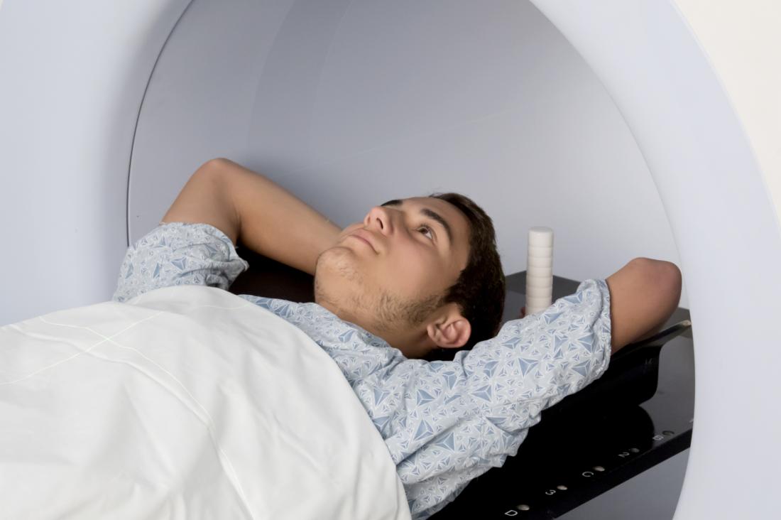
Radiation Therapy
The most common cause of mortality after a successful cancer resection is a return of RLS. Radiation therapy (RT) has long been an important part of the multimodal treatment of advanced or metastatic cancer with affected margins.
However, there has been a lot of discussion about whether RT should be used adjuvantly or neoadjuvantly based on what we've learned from extremity sarcomas. Surgical resection of an extremity soft tissue sarcoma is followed by radiation or brachytherapy, which improves local control but not survival.
Different RT doses and fractionations, concurrent chemotherapy use, various delivery modalities, various timings (pre, intra or post-surgical), as well as various energy carriers are all used to treat this particular cancer type in retroperitoneal sarcoma trials (photons, electrons, protons, or carbon ions).
Prior to surgery, radiotherapy is often preferred. Preoperative radiation shields the radiation-sensitive tissues and organs that fill the resection bed following a massive tumor excision. Patients are more likely to adhere to and tolerate their drug regimens prior to surgery. In healthy, well-perfused tissue, the biological effects of RT are more pronounced than in damaged tissue.

Systemic Therapy
Chemotherapy is used in the treatment of advanced or metastatic soft-tissue sarcomas. Anthracyclines such as doxorubicin, epirubicin, and ifosfamide are all effective. Gemcitabine, docetaxel, trabectedin, and pazopanib are viable alternatives for second or third-line treatment of resistant illnesses.
The histological type and grade of a tumor have an effect on the chemosensitivity of STS. For leiomyosarcoma, gemcitabine and docetaxel are the most effective treatments for angiosarcoma; for desmoid tumors, liposomal doxorubicin is the most effective treatment. The histological subtype and grade of liposarcoma affect its chemosensitivity.
Treatment outcomes for patients with liposarcoma were monitored by experts at the Royal Marsden Hospital. Patients with myxoid liposarcoma had a greater survival rate than those with other liposarcomas.
There was a 25% difference in response rates between well-differentiated and dedifferentiated liposarcomas. Liposarcoma of the upper and lower limbs reacted better than other types of liposarcoma.
Pretreatment Biopsy
In most cases, a preoperative biopsy is unnecessary because RLS patients are diagnosed based on their iconography. Patients with resectable retroperitoneal masses may not require a pre-treatment biopsy.
Depending on the patient, imaging may indicate another condition that does not require surgery (lymphoma, Ewing sarcoma, GIST). Pretreatment histology confirmation is required for those at risk of incomplete resection.
Compared to open or laparoscopic treatments, an image-guided core or small needle aspiration biopsy is a safe and effective alternative in these individuals.
Extent Of Surgery Versus Tumor Biology
Since there are large blood vessels, nerves, and bone structures surrounding the tumor, it is impossible to remove the tumor with a margin of normal tissue. It is estimated that three out of every four patients will die due to getting an ongoing stomach illness in their area of residence.
Several centers propose liberal compartmental, en-bloc excision as a means of reducing the risk of local recurrence. All liposarcomas have an amplification of 12q13-15, which results in the overexpression of MDM2 and CDK4. This is due to the major ontogenetic differences between normal subcutaneous and visceral fat tissue.
However, the illness's molecular biology is expected to differ between the limb and retroperitoneal disease locales because of this heterogeneous nature. Because high-grade, dedifferentiated tumors are more likely to return and spread, surgery is rarely an option for treating them.
People Also Ask
Where Is A Retroperitoneal Liposarcoma?
An uncommon malignant tumor of mesenchymal origin, retroperitoneal liposarcoma can occur in any fat-containing area of the body. It is a primary retroperitoneal neoplasm and is one of the most prevalent.
What Is The Most Commonly Associated With Retroperitoneal Sarcoma?
Adult retroperitoneal soft tissue sarcomas are found in a wide variety of forms, with some studies reporting higher rates than others. However, in most investigations, liposarcomas, leiomyosarcomas, and malignant fibrous histiocytomas are the most frequently seen cell types (MFH).
How Is Retroperitoneal Mass Treated?
Retroperitoneal tumors should be treated as soon as possible by a multidisciplinary team of surgeons with extensive experience in this area. Wide surgical excision is the only current therapeutic option that has been proven to extend the lives of individuals with these malignancies.
How Is Retroperitoneal Mass Diagnosed?
Radiology and histology are used to make the initial diagnosis of retroperitoneal tumors. In cases of stomach pain or distension, ultrasonography (US) is often the first test conducted in a gynecological consultation clinic.
Conclusion
Retroperitoneal liposarcoma is a rare cancer that typically goes undetected until it's too late because of its lack of early symptoms. Staging and evaluating the disease correctly directs the therapeutic plan.
To ensure a long-term, cancer-free existence, patients who have macroscopically full resection are best served. It's still unclear how disease biology determines the extent of "compartmental" resection in these people.
Prospective randomized trials are currently being conducted by the EORTC to determine whether preoperative radiation can lower the risk of local recurrence after cancer surgery. At the same time, researchers are uncovering the molecular mechanisms of sarcoma and developing tailored medications that could one day contribute to current systemic treatments.
Surgeons should only do a full surgical excision at specialized centers with a team of experts. When it comes to cancer recurrence and metastasis, a multidisciplinary team should be involved and potentially conduct a study.

James Pierce
Author

Karan Emery
Reviewer
Latest Articles
Popular Articles