Diabetic Nephropathy - Its Relationship With Cell Apoptosis
High glucose (HG) triggers apoptosis, which aids in the treatment of diabetic nephropathy. However, the mechanism by which HG triggers apoptosis is still unknown. Apoptosis plays a role in the development and prevention of a number of diseases. Humans benefit from apoptosis because it causes cells with damaged DNA to commit "suicide" before becoming cancerous.
Author:Katharine TateReviewer:Karan EmeryAug 04, 2022149 Shares2.4K Views

High glucose (HG) triggers apoptosis, which aids in the treatment of diabetic nephropathy. However, the mechanism by which HG triggers apoptosis is still unknown.
Apoptosis plays a role in the development and prevention of a number of diseases. Humans benefit from apoptosis because it causes cells with damaged DNA to commit "suicide" before becoming cancerous.
It also contributes to muscle atrophy, which occurs when the body decides that wasting calories on maintaining muscle cells that aren't used on a regular basis is no longer necessary.
What Is Apoptosis?
Apoptosis programs cell death in multicellular organisms. It's a tightly regulated process that occurs naturally during development and aging to maintain tissue cell populations.
In a developing embryo, apoptosis between the digits separates fingers and toes. Apoptosis occurs when cells are damaged by disease or toxins. Light microscopy shows cell shrinkage and pyknosis in early apoptosis.
Cell shrinkage densifies the cytoplasm and compacts the organelles. The most distinctive feature of apoptosis is pyknosis, caused by chromatin condensation. Eventually, apoptosis produces apoptotic bodies, which phagocytic cells engulf and remove before the contents cause inflammation.
Intrinsic pathways, in which a cell receives a signal to start apoptosis from its own genes or proteins in response to DNA damage, and extrinsic pathways are the two major apoptosis pathways. When a cell has outlived its usefulness, the extrinsic pathway is activated.
Two Pathways Of Apoptosis
Two pathways can trigger apoptosis:
- Intrinsic Pathway
- Extrinsic Pathway
The Extrinsic Pathway
External stimuli, like ligand binding at cell death receptors, activate this pathway, causing apoptosis. Receptors are in the Tumor Necrosis Factor (TNF) gene family. Caspases are activated by receptors.
The Intrinsic Pathway
This pathway triggers apoptosis by biochemical stress, DNA damage, and lack of growth factors. The pathway is regulated by Bax and Bcl-2. Molecular groups determine a cell's survival or apoptosis in response to stimuli.

Diabetic Nephropathy
Some Examples Of Apoptosis
From Tadpole To Frog
Frog tadpoles destroy and re-absorb entire body structures during metamorphosis.
Apoptosis signals cause gill, fin, and tail cells to die as the tadpole matures. Disassembled cells are used to build new limbs and feed them.
Human Nervous System Development
Apoptosis kills many cells during early nervous system development.
Scientists don't know why the developing nervous system has so much cell death. Some speculate that this is because forming the correct connections is a complex and difficult process for young neurons, and maximizing the nervous system's efficiency is in the organism's best interest.
Mouse Feet
Embryonic mouse feet are spade-shaped. Apoptosis splits the feet into five toes during development. To create toe gaps, connecting cells die off.
This shows how programmed cell death can be used to create useful structures and remove unwanted ones.
How Apoptosis Affects Cancer?
Apoptosis removes harmful cells from the body. A cell enters apoptosis when its DNA is damaged. DNA damage activates apoptosis pathways, preventing cancerous cell transformation.
Clearly, this method doesn't always work. All cancers result from a damaged cell failing to commit apoptosis and reproducing.
Damaged apoptosis genes may prevent cell death. Some doctors and scientists study apoptosis to trigger it in cancer cells with new medications or treatments.

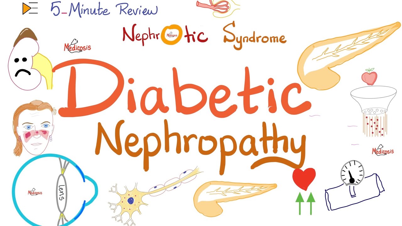
Diabetic Nephropathy - Nephrotic Syndrome - Kidney Pathology
Relationship Between Apoptosis And Diabetic Nephropathy
Because it can balance apoptosis, the tuberin/mTOR pathway in type 1 diabetes and in PTE cells exposed to HG was studied.
Diabetes-related kidney damage led to more apoptotic cells in diabetic rodents. Diabetes enlistment increases tuberin phosphorylation in relation to mTOR initiation (measured by p70S6K phosphorylation), Bcl-2 inactivation, cytosolic cytochrome c articulation, caspase 3 activation, and PARP cleavage; insulin treatment prevents these changes. In vitro exposure to HG increased tuberin and p70S6K phosphorylation, Bcl-2 phosphorylation, cytosolic cytochrome c articulation, and caspase 3 activity.
High glucose moved the caspase substrate YY1 to the core, which improved PARP cleavage. The mTOR inhibitor rapamycin reduced the number of apoptotic cells induced by HG and the downstream effects of mTOR activation. SiRNA reduced PARP cleavage by silencing tuberin.
The tuberin/mTOR pathway promotes apoptosis in rounded epithelial cells in diabetes, with PARP cleavage by YY1 interfering. Biotin is a water-soluble nutrient that aids in starch, fat, and amino acid digestion. Named for its role as a fundamental cofactor for five carboxylases.
The mitochondrial carboxylase propionyl-CoA carboxylase (PCC) uses biotin. Reduced PCC action in blood lymphocytes reflects intracellular biotin deficiency in trial biotin deficiency patients.
PCC enactment coefficient (AC), which compares PCC action after hatching unblemished, suitable blood lymphocytes with biotin to PCC action before brooding with biotin, is another measure of tissue biotin status. Biotin transport into lymphocytes increases apo-PCC mass by increasing PCC AC. During peripheral biotin deficiency, AC expands, mirroring intracellular apo-PCC. Due to its small size (244.31 Daltons) and proximity to the watery stage, hemodialysis and peritoneal dialysis likely drain biotin.
Dialysis patients are given 150-300 g of biotin orally daily, which is 5-10 times the AI for healthy adults. Poor biotin status may contribute to RLS pathogenesis in some dialysis patients. Background research included ESRD patients on dialysis.
Prior to enlistment, members' primary care doctors assessed RLS based on clinical side effects, including a desire or need to move the legs, which is often accompanied or caused by loud noises, side effects are more severe when lying still, manifestations are at least halfway and incidentally diminished by action, and side effects show a circadian pattern, with a peak in the evening or around evening.
At the time of enlistment, all members with RLS had an IRLS Group rating scale score of at least 4. This scoring system is dependable and legitimate. The IRLS examiner uses a 10-point scale to assess sleep disturbances and anxious leg symptoms.
People Also Ask
How Does Diabetes Cause Cell Death?
Hyperglycemia is a hallmark of diabetes, and although managing this complication can slow but not halt the progression of the disease's complications, it cannot stop them entirely. A high blood sugar level can set off a number of metabolic signaling pathways, which can ultimately result in inflammation, the secretion of cytokines, cell death, and the development of diabetic complications.
Does Insulin Increase Apoptosis?
Insulin has the ability to prevent apoptosis in HepG2 cells by lowering the levels of oxidative stress. This is accomplished through the phosphatidylinositol 3-kinase and extracellular signal-regulated protein kinase-dependent signaling pathways.
How Does Hyperglycemia Cause Apoptosis?
Based on these findings, it appears that hyperglycemia directly induces the death of myocardial cells via the apoptotic pathway in vivo. Apoptosis of the myocardium caused by hyperglycemia is mediated, at least in part, by activation of the cytochrome c-activated caspase-3 pathway. This pathway's activation may be triggered by reactive oxygen species (ROS) that are produced when high levels of glucose are present.
What Happens To Cells In Hyperglycemia?
When there is an increase in the amount of glucose in your blood, it sends a signal to your pancreas to secrete insulin. Your cells are unlocked by the insulin, allowing glucose to enter and provide the fuel that is necessary for your cells to function properly. Any glucose that is not used is converted into glycogen and stored in your liver and muscles.
Conclusion
Apoptosis is triggered by physiologic and pathological stimuli. Not all cells respond to the same stimulus equally. Irradiation or chemotherapy drugs can damage DNA, triggering apoptosis in some cells via a p53-dependent pathway. Apoptosis' impact on diabetic nephropathy is being studied.
Separating apoptosis and necrosis, which can occur simultaneously, sequentially, or independently, is difficult. The type and/or level of stimulation can affect whether cells die by apoptosis or necrosis. Low doses of heat, radiation, hypoxia, and cytotoxic anti-cancer drugs can cause apoptosis, but high doses can cause necrosis.

Katharine Tate
Author

Karan Emery
Reviewer
Latest Articles
Popular Articles
