Traumatic Brain Injury - The Role Of Markers In Inflammation
The term "secret epidemic" has been used to describe traumatic brain injury (TBI) for many years. A significant inflammatory response is produced in the wounded brain minutes after a traumatic impact. Even mild traumatic brain injuries can cause cognitive deficiencies, focus issues, tiredness, and headaches. Many survivors of traumatic brain injuries are left permanently disabled.
Author:Katharine TateReviewer:Karan EmeryOct 11, 2022117 Shares1.6K Views

The term "secret epidemic" has been used to describe traumatic brain injury(TBI) for many years.
A significant inflammatory response is produced in the wounded brain minutes after a traumatic impact.
Even mild traumatic brain injuries can cause cognitive deficiencies, focus issues, tiredness, and headaches.
Many survivors of traumatic brain injuries are left permanently disabled.
This post-traumatic scream has a complicated biological makeup that includes the activation of local glial cells, microglia, and astrocytes as well as the invasion of blood leukocytes.
Currently, the main goals of TBI patient care are the monitoring and maintenance of normal intracranial pressure (ICP) and cerebral perfusion pressure (CPP).
Treatment and care of a TBI patient may involve reducing ICP in addition to other variables.
What Is Traumatic Brain Injury?
Anyone can sustain a traumatic brain injury (TBI) after being hit in the head.
Gunshot wounds and non-penetrating injuries, such as being hit in the head in a car accident, are both examples of penetrating injuries.
The degree of a traumatic brain injury varies. TBIs can cause temporary or permanent brain damage, or even death in the most severe cases.
TBIs can affect anyone, although men are more likely than womento sustain one. A higher percentage of adults over the age of 65 are affected by TBIs.
People in this age range are more likely to trip, fall, and injure their brains.
However, even infants can sustain a traumatic brain injury (TBI) if they fall from a bed or changing table, or if they are abused.
TBIs are more common in certain occupations or activities than in others.
What Are Some Of The Symptoms Of A Traumatic Brain Injury?
The signs and symptoms of a traumatic brain injury (TBI) can vary in severity. Passing unconscious after taking a hit is an important telltale indication.
For a few minutes, some people are dazed; for longer periods, they are unresponsive (coma or persistent vegetative state).
Mild traumatic brain injuries (TBIs) can cause a wide range of symptoms, the majority of which appear within hours or days of the trauma.
The intensity of a symptom may not become apparent until a person returns to school or a job after an absence.
TBI symptoms include:
- Mood or behavior shifts.
- Memory lapses or a lack of focus.
- Seizures and convulsions.
- Blurred eyesight or dilation of the pupils.
- Unsteadiness, faintness, or exhaustion.
- Headaches.
- Vomiting and diarrhoea.
- Anxiety or restlessness.
- Light and odor sensitivity.
- Sleeping excessively or insufficiently.
- Speech that is slurred.
TBIs in infants and children may also include:
- Become hysterical and weep uncontrollably.
- Refuse to consume any food or fluids, including breastmilk.
What Are The Types Of Traumatic Brain Injuries?
The severity of a head injury is judged by a number of factors, including loss of consciousness, certain neurological symptoms that occurred at the time of the event, loss of memory of the injury and the time surrounding it, and abnormalities on a head CT or brain MRI.
TBI comes in a variety of forms and severity levels:
Mild concussion (mTBI):TBIs caused by concussions are by far the most prevalent. Concussions account for three out of every four cases of TBI. A "dazed" or "disoriented" state, lasting less than 30 minutes, might be one of the symptoms of a mild traumatic brain injury (mTBI). People who have had a mild traumatic brain injury (mTBI) may have disorientation for up to one day, which is distinct from memory or concentration issues.
Moderate TBI:This form of lesion to the brain results in a prolonged period of unconsciousness, usually lasting more than 30 minutes but not exceeding a day. Up to a week of disorientation is possible.
Severe TBI:For more than a full day, people with this form of brain injury are unconscious. A CT or MRI scan of the head is often used to diagnose these injuries.
Uncomplicated TBI:Regardless of mild, moderate, or severe grade, head CT or brain MRI are all normal.
Complicated TBI:A brain MRI or CT scan will reveal abnormalities, such as hemorrhages, in the brain.
Closed:The vast majority of traumatic brain injuries (TBIs) have been successfully treated. A closed traumatic brain injury (TBI) is one in which the brain has been struck but the force has not penetrated the skull. The brain swells as a result of this hit, which injures it.
Open:Healthcare providers may refer to an open TBI as a penetrating TBI. A bullet, knife, or other object passing through the skull causes this damage. Direct injury to brain tissue occurs if the item enters the brain.
Nontraumatic:A hypoxic/anoxic brain damage is also known. Other causes of traumatic brain injury Events like choking and nearly fatal drownings can lead to strokes. An oxygen deprivation in the brain can result from these events (cerebral hypoxia).

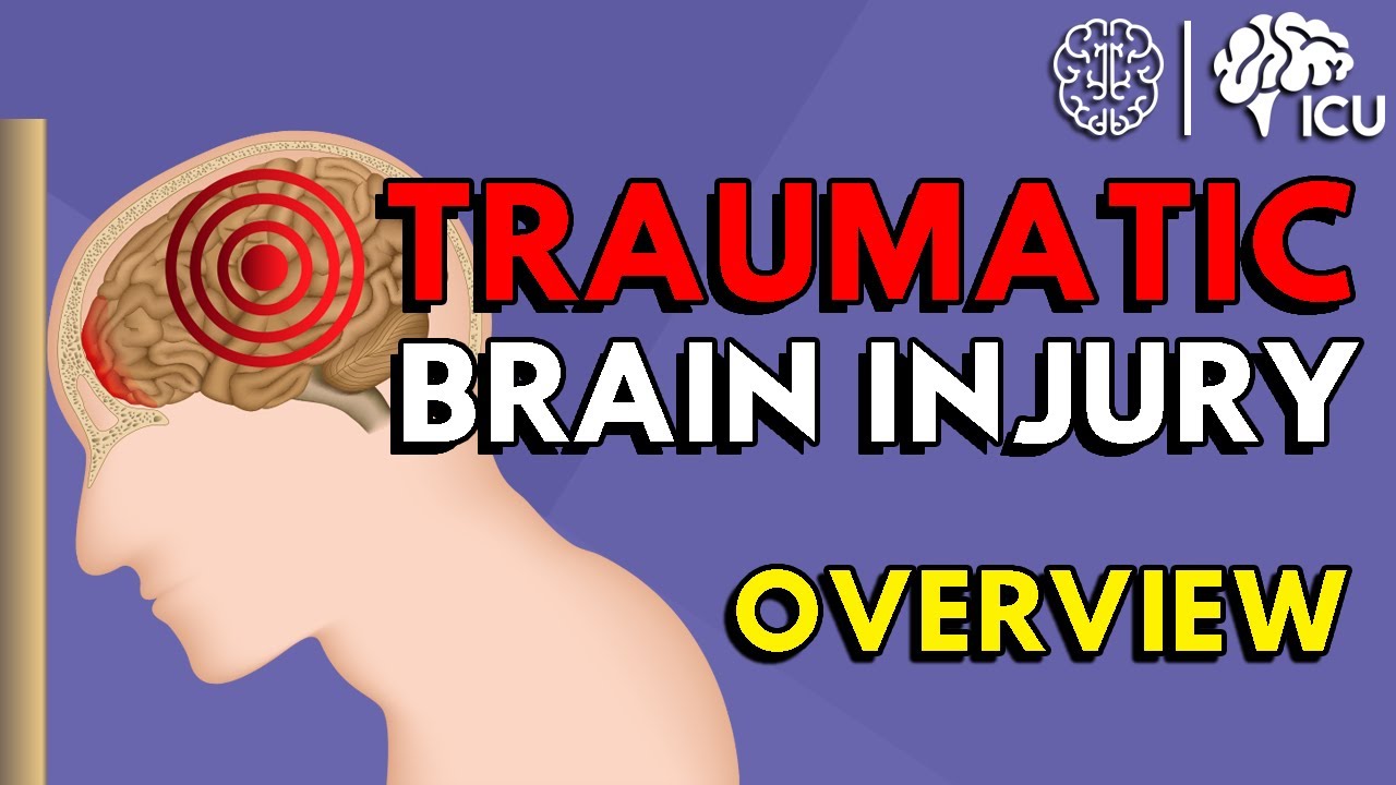
Overview of Traumatic Brain Injury (TBI)
The Development Of Biomarkers In TBI
Tissue damage can be detected by S100B and NSE, as well as by MBP, which is a marker of myelin basic protein (MBP).
Several observational clinical TBI studies have found a connection between S100B and the severity of the initial brain injury (GCS), the size of the brain damage (on CT/MRI scans), and the neurological outcome (Glasgow Outcome Scale/Extended; GOSE).
Only lately have biomarkers been employed in clinical trials that collect prospective data, analyze results, and evaluate the effectiveness of interventions.
This study aims to demonstrate the link between neuroprotection and the lowering of biomarkers in TBI patients.
Six-month neurological ratings are not as sensitive as biomarkers in predicting prognosis early on.
Due to the small number of patients in clinical studies, biomarkers may be a better way to find variations in TBI patient outcomes.
S100B, a low-molecular-weight calcium-binding astrocyte protein, is the most studied biomarker for TBI.
As a TBI biomarker, the usefulness of S100B is predicated on the fact that after a brain injury, its normally modest levels of serum and CSF expression are rapidly released.
Injury severity and a poor prognosis are both associated with high levels of S100B expression.
A biomarker for TBI that doesn't pass the blood-brain barrier (BBB) and doesn't always predict prognosis isn't acceptable for S100B.
Numerous molecules have been examined in the search for a TBI biomarker, including GFAP, NSE, MBP, II-spectrin breakdown products (BDPs), ubiquitin C-terminal hydrolase-L1, and other cytokines.
The Potential Of Inflammatory Cytokines As Biomarkers Of TBI
The inflammatory response to TBI is modulated by pro and anti-inflammatory cytokines. Cytokines are produced by leukocytes and glial cells.
A quarantine is quickly instituted following a TBI. A complex network of inflammatory mediators is formed by numerous overlapping cytokines.
Anti-inflammatory cytokines counteract the pro-inflammatory effects of cytokines that stimulate inflammation.
After a brain injury, the expression profile of each cytokine can be analyzed to see how much tissue damage has occurred.
In rodents and humans, the cytokine levels differ by tissue or fluid (brain tissue, cerebrospinal fluid, serum, plasma), and the temporal profile of the inflammatory response in the brain varies widely from one species to the next.
Biomarkers and cytokines for brain damage are listed below.
Interleukin-1
One of the most important groups of cytokines in the inflammatory cascade is the interleukin-1 (IL-1) family.
One of the most well-known molecules in relation to acute TBI is IL-1, which has been extensively researched in both focal and diffuse injury models.
Numerous cell types in the brain express the IL-1 receptor type I (IL-1R), which is hypothesized to be a mediator of many of the effects of IL-1 cytokines.
Some of the effects of IL-1 cytokines may not be dependent on the IL-1R, however.
Some of these actions may be due to IL-1's ability to influence gene transcription and RNA splicing in the nucleus.
There are four members of the IL-1 family, all of which are connected to each other: the agonist IL-1α and IL-1β, the antagonist IL-1ra, and agonist IL-18.
TBI patients are more likely to report the IL-1β isoform than any of the others.
It has been shown that the release of PLA2, prostaglandins, and cyclooxygenase-2 are all aided by the pro-inflammatory cytokine IL-1β (COX-2).
In addition, IL-1β is thought to work primarily by controlling the release of other cytokines.
The apoptotic adherence of leukocytes to endothelial cells, the breakdown of the BBB, and the production of edema have all been linked to IL-1.
Interleukin-10
Since it inhibits the synthesis of a wide range of pro-inflammatory mediators, including IL-1β and TNF, as well as IL-1α, GM-CSF, IL-6, IL-8, IL-12, and IL-18, interleukin-10 is regarded to be primarily an anti-inflammatory cytokine.
Most importantly, it inhibits IL-1β and TNF, two cytokines that have been linked to the onset and propagation of an inflammatory response.
After LFP injury in rats, IL-10 therapy resulted in improved outcomes and lower levels of IL-1β and TNF in the brain tissues.
Interleukin-6
Scientists have examined interleukin-6 extensively and discovered that it is involved in numerous physiological processes, both beneficial and pathological.
Inflammation, immunology, bone metabolism, hematopoiesis, and nerve cell formation are all under the influence of IL-6.
Alzheimer's, and brain trauma are all linked to IL-6, as are aging and osteoporosis.
IL-6 is not only prevalent in the central nervous system (CNS), but it is also markedly increased in response to a brain injury.
Other Cytokines
TBI biomarkers are not limited to the cytokines outlined above; there are many other immune modulators that could be beneficial in the diagnosis and treatment of this condition.
Additionally, GM-CSF, which is produced by CNS neurons, astrocytes, and microglia, is a possible biomarker.
CSF contains GM-CSF, which is released by vascular endothelial cells and travels over the BBB.
Post-mortem tissue from TBI patients who died between 6 and 122 hours after the injury showed a significant increase in this protein.
For the first time, we have discovered that GM-CSF is more significantly expressed in the CSF of patients with secondary hypoxia compared to normoxic TBI patients, and also in diffuse TBI versus localized TBI (unpublished results).
This cytokine has different levels of expression in different injury types, which warrants additional exploration.
P43/pro-EMAP II, which is a pro-inflammatory cytokine associated with apoptosis, may also be of interest in this study.
Only the cytokine p43/pro-EMAP II was found to be differentially expressed in rats 24 hours after ischemic injury versus penetrating TBI.

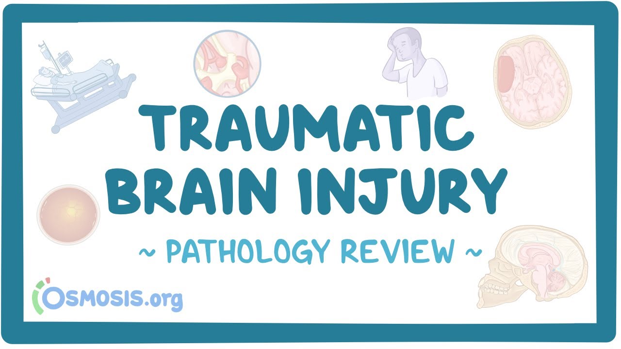
Traumatic brain injury: pathology review
Other Markers Of Inflammation
A few additional substances, including cytokines, may serve as biomarkers for brain injury.
For instance, IL-6 regulates GFAP production by activating the JAK/STAT pathway. It is well-known that GFAP expression in serum increases following TBI.
TBI survivors with high concentrations of GFAP in their blood are more likely to die than those with lower amounts of the protein.
Within four hours of the event, TBI severity (GCS) and CT lesions have been connected to the blood GFAP-BDPs of individuals with mild and severe TBI.
The IMPACT Outcome Calculator and CSF and serum GFAP values were used in a recent study to acquire a clearer sense of what would happen.
Another finding was that in the cerebrospinal fluid (CSF), there was a substantial connection between the expression of SBDP145 (a breakdown product of II-spectrin) and death at 6 months.
People Also Ask
How Is Traumatic Brain Injury Diagnosed?
Diagnosis of TBI
As part of the evaluation process, patients often undergo a neurological exam.
This test measures a person's cognitive abilities as well as their motor and sensory abilities, including their eye movement and reflexes.
All TBIs cannot be detected by imaging techniques such as CT scans and MRI scans.
Who Is Most At Risk Of Traumatic Brain Injury?
If you're between the ages of 15 and 19, you're the third most likely age group to suffer a traumatic brain injury that requires hospitalization.
Is Traumatic Brain Injury Permanent?
There is hope for functional recovery despite the fact that traumatic brain injury damage is irreversible since injured brain cells cannot renew or repair themselves.
This is due to the fact that healthy brain cells may be able to reorganize and improve functions that are damaged by TBI.
Is A Traumatic Brain Injury A Concussion?
A bump, blow, or jolt to the head or a strike to the body that causes the head and brain to move rapidly back and forth creates a concussion, a type of traumatic brain injury (TBI).
Conclusion
Nerve injury biomarkers have the potential to improve the diagnosis and treatment of patients.
Moreover, they may be able to help develop new treatments.
The inflammatory response that occurs when a patient is suffering from traumatic brain injury could provide doctors with a multitude of markers that reveal precise information about the lesion.
According to studies on animal models of traumatic brain injury, the brain produces a lot of inflammatory mediators following an injury.
Many of these change their expression quickly, reaching a thousand times their normal levels within hours of being harmed.
We may be able to learn more about a person's damage and the degree of complexity involved when several injuries occur from the amount, timing, and duration that these mediators are active.
More precise treatments for secondary injury may be made possible if these cytokines are better understood and their functions in the process are better understood.
It may also be possible to track the impact of future pharmacological treatments by measuring these cytokines.
SCI is treated with methylprednisolone (MP), which has anti-oxidant and anti-inflammatory properties.
Jump to
What Is Traumatic Brain Injury?
What Are Some Of The Symptoms Of A Traumatic Brain Injury?
What Are The Types Of Traumatic Brain Injuries?
The Development Of Biomarkers In TBI
The Potential Of Inflammatory Cytokines As Biomarkers Of TBI
Interleukin-1
Interleukin-10
Interleukin-6
Other Cytokines
Other Markers Of Inflammation
People Also Ask
Conclusion

Katharine Tate
Author

Karan Emery
Reviewer
Latest Articles
Popular Articles
